Vascular Ultrasound Specialist in Michigan
CardioQ – Heart & Wellness Center, a leading healthcare provider in Michigan, offers specialized diagnostic services including Duplex Doppler Ultrasound or Vascular Ultrasound performed by our expert Vascular Ultrasound specialists.
Our specialists are highly trained and experienced in using advanced ultrasound technology to assess blood flow and detect abnormalities in the arteries and veins throughout the body. With a commitment to providing compassionate care and accurate diagnoses, our Vascular Ultrasound specialists work closely with patients to develop individualized treatment plans that meet their unique needs.
At CardioQ, we strive to provide the highest quality care to help our patients achieve optimal vascular health.
Vascular Ultrasound or Duplex Doppler Ultrasound
Vascular ultrasound, also known as a Doppler ultrasound, is a non-invasive diagnostic imaging test that uses high-frequency sound waves to evaluate the blood flow in the arteries and veins of the body. It is a painless, safe, and effective way to detect abnormalities in blood vessels and diagnose a variety of vascular conditions, including deep vein thrombosis, peripheral artery disease, and carotid artery disease.
During a vascular ultrasound, a specialized ultrasound machine is used to produce images of the blood vessels and measure the speed and direction of blood flow. The images and measurements obtained can help identify areas of blockage or narrowing in the blood vessels, as well as detect blood clots or other abnormalities that may be affecting blood flow.
Vascular ultrasound is a non-invasive alternative to other diagnostic tests, such as angiography, which require injection of contrast material into the blood vessels or invasive procedures like catheterization. It is a relatively quick and painless procedure that can be performed in a healthcare provider’s office or a hospital setting, and does not require any special preparation.
Overall, vascular ultrasound is a safe and effective tool for diagnosing and monitoring a wide range of vascular conditions.
Time it Takes
The length of time it takes to perform a vascular ultrasound can vary depending on the area being examined and the complexity of the case. Typically, a vascular ultrasound can take anywhere from 30 minutes to an hour to complete.
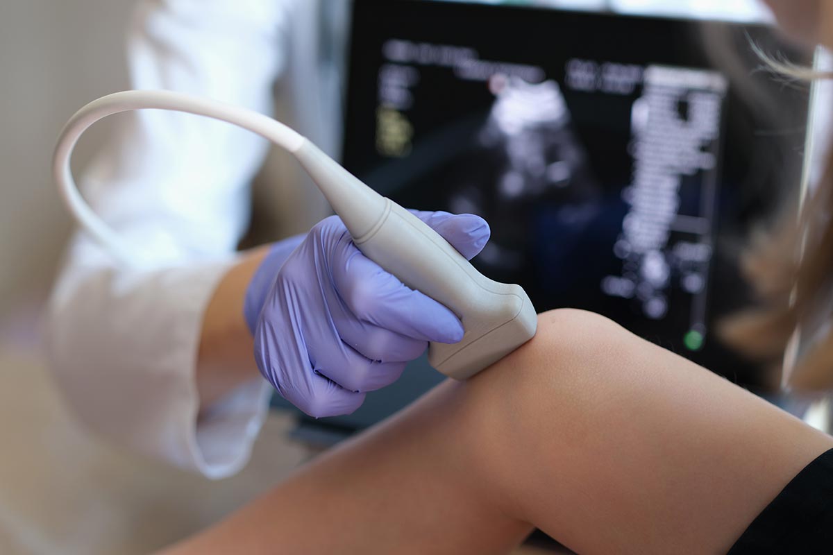
During the procedure, the patient lies on a table while a technician or vascular ultrasound specialist applies a special gel to the skin over the area being examined. The gel helps to transmit the sound waves from the ultrasound machine into the body and produce clear images of the blood vessels.
The technician then moves a handheld device called a transducer over the skin to capture images of the blood vessels from different angles. The images are displayed on a monitor in real-time, allowing the technician or specialist to evaluate blood flow and detect any abnormalities.
After the procedure is complete, the patient can typically resume normal activities immediately. The results of the vascular ultrasound are usually interpreted by a vascular specialist and communicated to the patient’s healthcare provider.
Getting the Results
After a vascular ultrasound, the images and measurements obtained are interpreted by a vascular specialist. The cardiologist will then review the results with the patient and discuss any abnormalities or concerns that were identified.
In some cases, the provider may order additional tests or procedures based on the results of the vascular ultrasound. For example, if a blockage or narrowing is identified in the blood vessels, the provider may recommend further imaging tests for further evaluation and treatment.
The results of a vascular ultrasound can provide valuable information about the health of the patient’s blood vessels and help diagnose a wide range of vascular conditions. With early detection and appropriate treatment, many vascular conditions can be effectively managed and even reversed, helping patients achieve better vascular health and overall well-being.
Common questions
Some common reasons why a healthcare provider may order a vascular ultrasound include:
- Peripheral artery disease (PAD): A vascular ultrasound can help diagnose PAD, a condition in which the arteries in the legs become narrowed or blocked, leading to decreased blood flow to the legs and feet.
- Deep vein thrombosis (DVT): A vascular ultrasound can detect blood clots in the veins of the legs or arms, a condition known as DVT, which can be life-threatening if left untreated.
- Carotid artery disease: A vascular ultrasound can assess the health of the carotid arteries in the neck, which supply blood to the brain. Narrowing or blockage of these arteries can increase the risk of stroke.
- Varicose veins: A vascular ultrasound can help diagnose varicose veins, a condition in which the veins in the legs become enlarged and twisted, leading to pain, swelling, and other symptoms.
- Follow-up after surgery: A vascular ultrasound may be recommended after surgery to evaluate the healing process and ensure that blood flow is returning to normal.
Overall, a vascular ultrasound is a safe and effective tool for diagnosing and monitoring a wide range of vascular conditions. If you have concerns about your vascular health or have been experiencing symptoms such as leg pain, swelling, or numbness, talk to your healthcare provider about whether a vascular ultrasound may be appropriate for you.
There are several different types of vascular ultrasound tests that can be performed depending on the specific area of the body being examined and the type of information needed. Some of the most common types of vascular ultrasound tests include:
- Carotid ultrasound: This type of vascular ultrasound examines the carotid arteries in the neck to assess blood flow to the brain and detect any narrowing or blockages that could increase the risk of stroke.
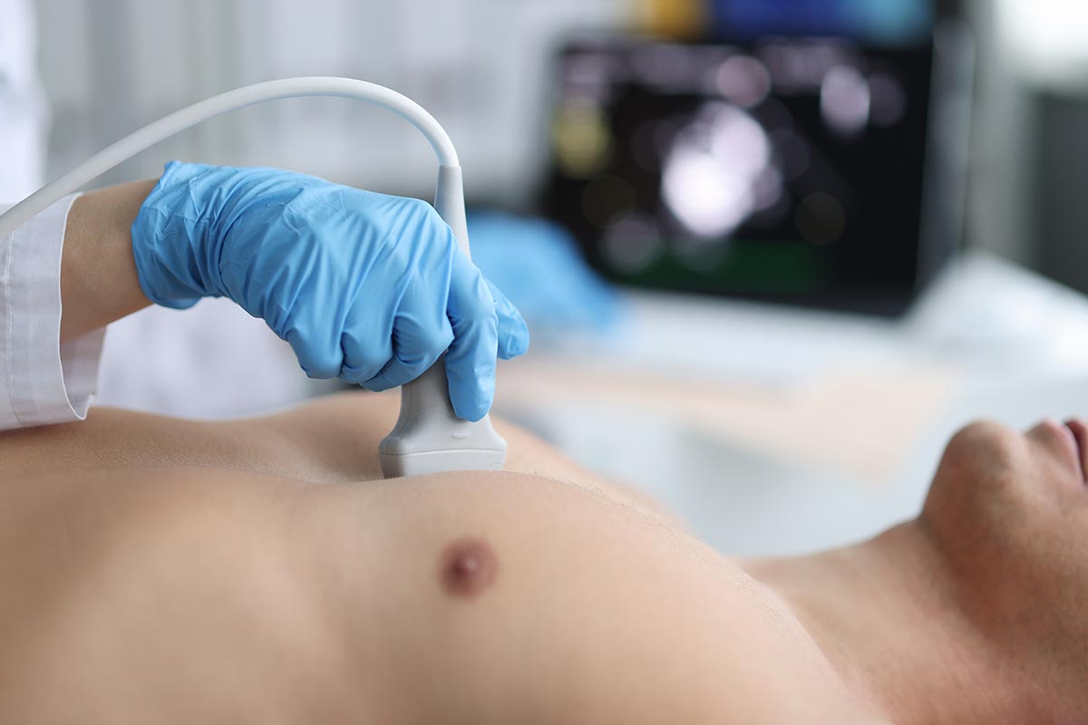
- Abdominal ultrasound: An abdominal ultrasound examines the blood vessels in the abdomen, including the aorta and its branches, to evaluate blood flow and detect any abnormalities.
- Renal artery ultrasound: This type of vascular ultrasound examines the blood vessels that supply the kidneys to evaluate blood flow and detect any narrowing or blockages that could lead to kidney damage.
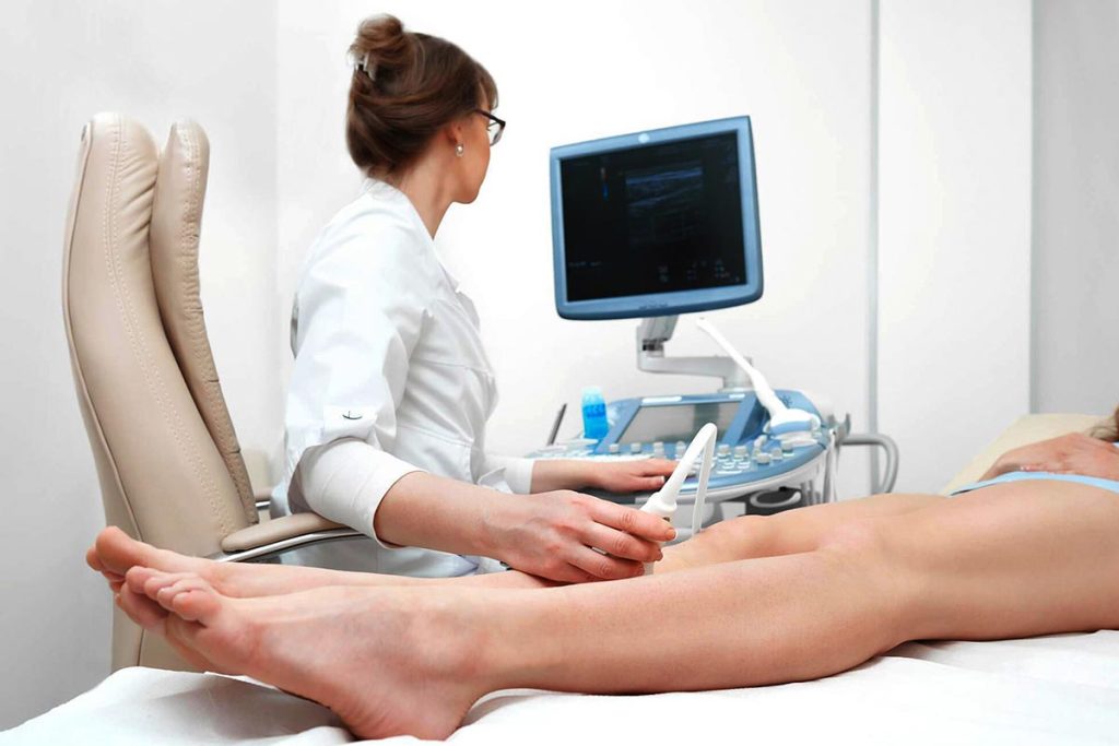
- Venous ultrasound: A venous ultrasound examines the veins in the legs, arms, or other parts of the body to detect blood clots, assess blood flow, and evaluate for conditions such as varicose veins.
- Arterial ultrasound: An arterial ultrasound examines the arteries in the legs, arms, or other parts of the body to evaluate blood flow and detect any narrowing or blockages that could lead to conditions such as peripheral artery disease.
These are just a few examples of the different types of vascular ultrasound tests that may be performed. The specific type of ultrasound recommended will depend on the individual patient’s symptoms, medical history, and the area of the body being examined.
During a vascular ultrasound, a specialist known as a vascular sonographer will use a small handheld device called a transducer to emit high-frequency sound waves into the body. These sound waves bounce off the blood vessels and are then picked up by the transducer, which creates detailed images of the blood vessels on a computer screen.
The vascular sonographer will typically begin by applying a gel to the skin over the area being examined. This helps to improve contact between the skin and the transducer and allows for better transmission of the sound waves.
The transducer is then moved back and forth over the skin, sending sound waves into the body and receiving the echoes that bounce back. The sound waves are processed by a computer to create detailed, real-time images of the blood vessels.
As the ultrasound is being performed, the vascular sonographer will be able to see the images on a computer screen and may adjust the position of the transducer or ask the patient to change positions to obtain better images.
The entire process typically takes between 30 and 60 minutes to complete, depending on the area of the body being examined and the specific information needed.
After the vascular ultrasound is complete, a radiologist or vascular specialist will interpret the images and provide a report to the patient’s healthcare provider. The provider will then review the results with the patient and discuss any abnormalities or concerns that were identified.
The results of a vascular ultrasound will depend on the specific type of ultrasound that was performed and the area of the body that was examined. However, in general, the results of a vascular ultrasound will provide information about the blood flow and condition of the blood vessels in the area being examined.
For example, if a carotid ultrasound was performed, the results may indicate the presence of plaque buildup in the carotid arteries, which can increase the risk of stroke.
If an abdominal ultrasound was performed, the results may indicate the presence of an abdominal aortic aneurysm or blockages in the renal arteries that could lead to kidney damage.
The results of a vascular ultrasound may also include measurements of blood flow velocity or pressure, which can help to diagnose conditions such as peripheral artery disease or deep vein thrombosis.
It is important to note that the interpretation of vascular ultrasound results requires specialized training and expertise. A cardiologist or vascular specialist will typically review the results of the ultrasound and provide a report to the patient’s healthcare provider, who will then review the results with the patient and recommend any necessary follow-up testing or treatment.